 E-submission
Articles
- Page Path
- HOME > J Mov Disord > Volume 5(1); 2012 > Article
-
Case Report
Apparently Ipsilateral Parkinsonism in a Patient with Chronic Subdural Hematoma - Tae Hwan Roha*, Dokyung Leea*, Il Ki Hongb, Deog Yoon Kimb, Tae-Beom Ahna
-
Journal of Movement Disorders 2012;5(1):18-20.
DOI: https://doi.org/10.14802/jmd.12005
Published online: April 30, 2012
aDepartments of Neurology, School of Medicine, Kyung Hee University, Seoul, Korea
bNuclear Medicine, School of Medicine, Kyung Hee University, Seoul, Korea
- Corresponding author: Tae-Beom Ahn, MD, PhD Department of Neurology, School of Medicine, Kyung Hee University, 23 Kyungheedae-ro, Dongdaemun-gu, Seoul 130-702, Korea, Tel +82-2-958-8448, Fax +82-2-958-8490, E-mail ricash@hanmail.net
-
*
These authors contributed equally to this work.
The authors have no financial conflicts of interest.
• Received: April 1, 2012 • Revised: April 25, 2012 • Accepted: April 25, 2012
Copyright © 2012 The Korean Movement Disorder Society
This is an Open Access article distributed under the terms of the Creative Commons Attribution Non-Commercial License (http://creativecommons.org/licenses/by-nc/3.0/) which permits unrestricted non-commercial use, distribution, and reproduction in any medium, provided the original work is properly cited.
- 7,468 Views
- 69 Download
- 1 Crossref
ABSTRACT
- Symptomatic parkinsonism secondary to ipsilateral lesion is rarely reported. Although the contribution of the contralateral lesions was assumed in some cases, the pathomechanism remains undetermined. Herein we report a patient with a subdural hematoma, who developed parkinsonism in the ipsilateral hemibody. Structural and functional imaging suggests the contralateral dopaminergic dysfunction as the major culprit of apparently ipsilateral parkinsonism.
- A 77-year-old man was admitted for memory loss, clumsiness of the right hand, and gait disturbance. He experienced headache and nausea 4 years before without any history of head trauma, which subsided without specific treatment. His right hand became clumsy around the episode. He had a difficulty in arising from the chair, and began to walk slowly. His symptoms were gradually aggravated afterward.
- A neurologic examination showed impaired orientation and memory recall with a 17/30 on Mini-mental status examination. Motor and sensory examination was normal. Deep tendon reflex (DTR) slightly increased in the right knee. His right hand showed a dystonic posture resembling a striatal hand deformity with intermittent resting tremor. Cogwheel rigidity was present in the right arm. He showed a stooped posture and impaired postural reflex. His right leg was less agile with mild external rotation of the foot and with mild shuffling.
- Brain MRIs were taken four years before and at admission. Both showed a huge SDH with hydrocephalus without significant interval change. The striatum (STR) was compressed and displaced, more severe on the left side (Figure 1). The compressed thalamus and STR on the right side was displaced downwardly and medially and abutted the right midbrain (MB) (Figure 1A and B, arrows). Dorsal MB lost its convexity with widened ventricle (Figure 1C).
- To investigate the nigrostriatal dopaminergic system, positron emission tomography with 18F-fluorinated N-3-fluoropropyl-2-beta-carboxymethoxy-3-beta-(4-iodophenyl) nortropane (18F-FPCIT) was done. 18F-FPCIT uptake was decreased in both STR, more severe on the left side, and similar in both MB. Dopaminergic innervation of the putamen was more decreased in the posterior portion, creating an anterior-to-posterior gradient (Figure 2).
- Surgical treatment was waived. His cognitive function was improved with donepezil. Levodopa treatment was mildly effective for his parkinsonism.
Case
- Subdural hematoma could be hardly considered as the primary diagnosis because of protracted course, slow progression, and the scarce signs of increased intracranial pressure. However, fixed dystonia, increased DTR, and the spastic nature of his gait were suggestive of secondary parkinsonism.
- The decreased uptake of 18F-FPCIT in STR could be resulted from direct compression of STR or the disruption of the nigrostriatal pathways.4 As the caudate nuclei retained their convexity, disrupted nigrostriatal pathways may be more likely to be responsible for abnormal 18F-FPCIT uptake. The loss of dorsal convexity of MB would be compatible with the compromised nigrostriatal pathway by hydrocephalus.4 The anterior-to-posterior gradient of dopamine transporter scan could be a feature of pre-synaptic damage.5
- In our patient, asymmetric involvement of the contralateral nigrostriatal pathway may result in apparent ILP. It remains enigmatic the absence of parkinsonism in the contralateral side to SDH, in spite of STR abnormality with decreased 18F-FPCIT uptake.
Discussion
Figure 1.A brain magnetic resonance imaging shows a huge subdural hematoma with calcification over the right cerebral convexity with prominent hydrocephalus (A: axial, B: coronal, C: sagittal). The compressed right brain is displaced abutting the midbrain (arrow), while the left side of midbrain retains its normal contour.
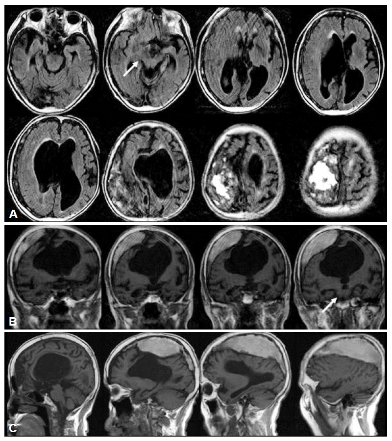
Figure 2.Positron emission tomography with 18F-fluorinated N-3-fluoropropyl-2-beta-carboxymethoxy-3-beta-(4-iodophenyl) nortropane (18F-FPCIT) shows the decreased uptake of 18F-FPCIT in both striatum, which is more severe on the left side (A: axial, B: coronal). In the midbrain, 18F-FPCIT uptake is well preserved on the left side (arrow). The 18F-FPCIT uptake of the right midbrain is preserved but appears as dispersed in the axial image (arrowhead) mixed with that of overlying thalamus (asterisk).

- 1. Polyzoidis KS, McQueen JD, Rajput AH, MacFadyen DJ. Parkinsonism as a manifestation of brain tumor. Surg Neurol 1985;23:59–63.ArticlePubMed
- 2. Kulali A, Tuğtekin M, Utkür Y, Erkurt S. Ipsilateral hemi-parkinsonism secondary to an astrocytoma. J Neurol Neurosurg Psychiatry 1991;54:653.ArticlePubMedPMC
- 3. Turjanski N, Pentland B, Lees AJ, Brooks DJ. Parkinsonism associated with acute intracranial hematomas: an [18F]dopa positron-emission tomography study. Mov Disord 1997;12:1035–1038.ArticlePubMed
- 4. Racette BA, Esper GJ, Antenor J, Black KJ, Burkey A, Moerlein SM, et al. Pathophysiology of parkinsonism due to hydrocephalus. J Neurol Neurosurg Psychiatry 2004;75:1617–1619.ArticlePubMedPMC
- 5. Im JH, Chung SJ, Kim JS, Lee MC. Differential patterns of dopamine transporter loss in the basal ganglia of progressive supranuclear palsy and Parkinson’s disease: analysis with [(123)I]IPT single photon emission computed tomography. J Neurol Sci 2006;244:103–109.ArticlePubMed
REFERENCES
Figure & Data
References
Citations
Citations to this article as recorded by 
- Secondary parkinsonism caused by chronic subdural hematomas owing to compressed cortex and a disturbed cortico–basal ganglia–thalamocortical circuit: illustrative case
Masao Fukumura, Sho Murase, Yuzo Kuroda, Kazutomo Nakazawa, Yasufumi Gon
Journal of Neurosurgery: Case Lessons.2021;[Epub] CrossRef
Comments on this article
 PubReader
 ePub Link
 Cite
Cite

