Skip Navigation
Skip to contents

JMD : Journal of Movement Disorders

OPEN ACCESS
SEARCH
Search

Articles

Page Path
HOME > J Mov Disord > Volume 11(1); 2018 > Article
Letter to the editor
Myotonia Congenita Can Be Mistaken as Paroxysmal Kinesigenic Dyskinesia
Aryun Kim1, Mihee Jang1, Han-Joon Kim1, Yoon Kim1, Dae-Seong Kim2, Jin-Hong Shin2, Beomseok Jeon1
Journal of Movement Disorders 2018;11(1):49-51.
DOI: https://doi.org/10.14802/jmd.17056
Published online: January 23, 2018

1Department of Neurology, Seoul National University Hospital, Seoul National University College of Medicine, Seoul, Korea

2Department of Neurology, Pusan National University Yangsan Hospital, Yangsan, Korea

Corresponding author: Beomseok Jeon, MD, PhD, Department of Neurology, Seoul National University Hospital, Seoul National University College of Medicine, 101 Daehak-ro, Jongno-gu, Seoul 03080, Korea / Tel: +82-2-2072-2876 / Fax: +82-2-3672-7553 / E-mail: brain@snu.ac.kr
• Received: September 7, 2017   • Revised: October 19, 2017   • Accepted: October 24, 2017

Copyright © 2018 The Korean Movement Disorder Society

This is an Open Access article distributed under the terms of the Creative Commons Attribution Non-Commercial License (http://creativecommons.org/licenses/by-nc/4.0/) which permits unrestricted non-commercial use, distribution, and reproduction in any medium, provided the original work is properly cited.

  • 7,309 Views
  • 128 Download
  • 4 Web of Science
  • 4 Crossref
Paroxysmal kinesigenic dyskinesia (PKD) is commonly thought to be first described by Kertez [1]. However, the typical clinical features of PKD were previously described by Shuzo Kure in a Japanese medical journal in 1892, as reviewed by Kato et al. [2]. The case described a 23-year-old male who had involuntary movement attacks with onset at 10 years of age that gradually increased in frequency. The attacks were triggered by sudden movement and started in the legs, sometimes spread to the body, and were very brief. They were preceded by a sensory aura, and the patient learned how to inhibit or stunt the attacks. He never lost consciousness, and abnormal neurological signs were not observed [2]. These descriptions are in perfect agreement with our current definition of PKD [3]. However, Kure misdiagnosed the case as an “atypical case of Thomsen disease [myotonia congenita (MC)],” despite noting that there were no long-lasting muscle contractions during the attacks and there was an absence of percussion myotonia [2].
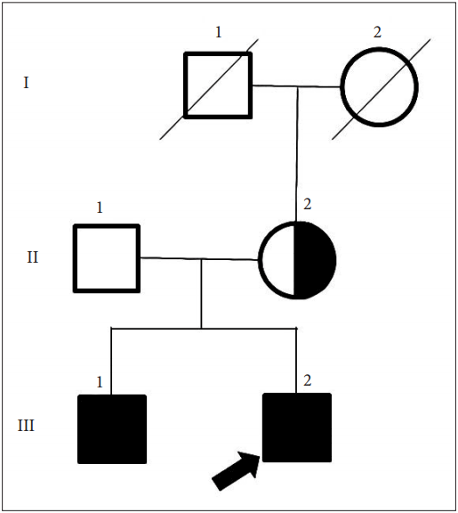
Herein, we describe a case of MC referred as PKD (Figure 1). MC and PKD are similar in that both are characterized by attacks of involuntary movement triggered by sudden movement and lasting only a brief duration. We will discuss the essentials for differential diagnosis between PKD and MC.
The pediatrics department referred a 19-year-old man (III-2) with possible paroxysmal movement disorder. Since 12 years old, he noted difficulty with initiating movement and getting stiff at the start of voluntary movements. For example, when getting on the bus, he had difficulty with the first couple of steps because of his feet getting curled up and becoming stiff, which got better after several steps. His hand use was similarly impaired and would become stiff upon first grip. These problems worsened after a period of prolonged rest but did not occur in the middle of active exercise. The patient was conscripted to the military after 7 years of stable conditions. In the army, he was observed as having the same problems, which interfered with his military training, and he was asked to seek a medical opinion. At first, he went to the pediatrician treating his elder brother (III-1), who had been diagnosed with MC due to a difficulty relaxing his hand grip since the age of 13. The patient’s brother (III-1) had myotonia when gripping his hand and percussion myotonia in the thenar muscles and tongue. The presence of myotonic discharge was confirmed using electromyography (EMG), and a diagnosis of MC was made without genetic analysis. The pediatrician considered the patient’s clinical history to be different from his brother’s and referred the proband (III-2) to us, believing he might have a different disease.
After a careful interview, it was determined that the involuntary movement attacks occurred simultaneously with immediate movement initiation, did not spread to other body parts, always consisted of stiffening without chorea, and were consistently triggered after a period of prolonged rest. Upon neurological examination, his cranial nerve, sensory, and motor systems were normal, apart from grip myotonia in both hands. He did not have percussion myotonia or muscular hypertrophy. In the clinic, a prolonged resting period was required before sudden standing and walking to induce muscle stiffening in the legs. This stiffening was localized to the legs and hindered his first several steps but eventually disappeared, and he was able to walk normally. His creatinine kinase level and nerve conduction tests were normal. However, EMG revealed myotonic discharge. His parents had no relevant clinical history and were both normal on neurological examination. With a clinical diagnosis of non-dystrophic myotonia, Sanger sequencing of CLCN1 (NM_000083.2, all exons and flanking introns) and SCN4A (NM_000334.4) was performed using genomic DNA from the proband and his family members. The proband (III-2), his elder brother (III-1), and his mother (II-2) all possessed a heterozygous missense mutation c.566C > G (p.S189C), and the patient was diagnosed with autosomal dominant MC (Thomsen disease) with a CLCN1 mutation. Treatment with 250 mg of acetazolamide twice a day reduced the frequency and severity of his symptoms to 20% of the initial state.
The involuntary movements in this case have several features of PKD: 1) they were triggered by sudden movements, 2) they were brief, 3) there was habituation, and 4) they occurred after periods of prolonged rest [4].
However, even without a family history of MC, there were still clues that pointed more towards MC than PKD [3,4]: 1) the involuntary movements began simultaneously with the trigger; 2) they were restricted to the body part where the triggering voluntary contraction occurred and did not spread to other body parts; 3) the movements were always stiffening without chorea; 4) they occurred “consistently” after prolonged rest; and 5) there was no aura.
Interestingly, there are 2 cases of CLCN1 c. 1205C > T mutations in a report of PRRT2-negative sporadic PKD in a Chinese study [5]. The cases were mistaken as PKD initially because the patients had childhood onset of paroxysmal stiffness of limbs triggered by sudden movement and accompanied by preserved consciousness during attacks. However, when examined after identification of CLCN1 mutation, both patients displayed clinical features of non-dystrophic myotonia. One patient felt aggravation in cold weather, and both showed an unsatisfactory response to carbamazepine.
In another report, there was co-existence of CLCN1 and PRRT2 mutations in the father and son of one family [6]. The son had seizures in early infancy and PKD from 11 years of age, and the presence of a PRRT2 mutation was determined by genetic analysis. However, carbamazepine 100 mg twice daily failed to improve his condition. Further questioning and examination illuminated the presence of more severe attacks in cold weather, relief after repetitive movements, and mild percussion myotonia. EMG showed myotonic discharge, and the CLCN1 gene study showed two heterozygous CLCN1 mutations: c.1723C > T (p.P575S) and c.2492A > G (p.Q831R). A family study showed that since the age of 15, the father had suffered from limb stiffness triggered by the initiation of movement. These symptoms worsened in cold weather, and a gene study of the father showed the same mutations in PRRT2 and CLCN1.
In summary, MC can be differentiated from PKD by confirming that symptoms do not spread to other body parts, there is an absence of any choreic movement or aura, and a consistent immediate occurrence from the triggering sudden movement is present.

Conflicts of Interest

The authors have no financial conflicts of interest.

B Jeon received a travel grant from Korea Research-Based Pharmaceutical Industry Association, Korean Pharmaceutical Manufacturers Association, Seoul National University, and Seoul National University Hospital. He also received research grants from the Ministry of Health and Welfare, Seoul National University Hospital, Sinyang Cultural Foundation, Korean Movement Disorder Society, Boryung pharm., Novartis Korea, Ipsen Korea, Samil Pharmaceuticals, Abbvie Korea, UCB Korea, Lundbeck Korea, and Sandoz Korea. Aryun Kim and Yoon Kim received a travel grant from the Korean Movement Disorder Society and Seoul National University Hospital.
Figure 1.
Pedigree of the family.
jmd-17056f1.gif
  • 1. Kertesz A. Paroxysmal kinesigenic choreoathetosis. An entity within the paroxysmal choreoathetosis syndrome. Description of 10 cases, including 1 autopsied. Neurology 1967;17:680–690.ArticlePubMed
  • 2. Kato N, Sadamatsu M, Kikuchi T, Niikawa N, Fukuyama Y. Paroxysmal kinesigenic choreoathetosis: from first discovery in 1892 to genetic linkage with benign familial infantile convulsions. Epilepsy Res 2006;70 Suppl 1:S174–S184.ArticlePubMed
  • 3. Bruno MK, Hallett M, Gwinn-Hardy K, Sorensen B, Considine E, Tucker S, et al. Clinical evaluation of idiopathic paroxysmal kinesigenic dyskinesia: new diagnostic criteria. Neurology 2004;63:2280–2287.ArticlePubMed
  • 4. Erro R, Sheerin UM, Bhatia KP. Paroxysmal dyskinesias revisited: a review of 500 genetically proven cases and a new classification. Mov Disord 2014;29:1108–1116.ArticlePubMed
  • 5. Wang HX, Li HF, Liu GL, Wen XD, Wu ZY. Mutation analysis of MR-1, SLC2A1, and CLCN1 in 28 PRRT2-negative paroxysmal kinesigenic dyskinesia patients. Chin Med J (Engl) 2016;129:1017–1021.ArticlePubMedPMC
  • 6. Li HF, Chen WJ, Ni W, Wu ZY. Paroxysmal kinesigenic dyskinesia and myotonia congenita in the same family: coexistence of a PRRT2 mutation and two CLCN1 mutations. Neurosci Bull 2014;30:1010–1016.ArticlePubMedPMC

Figure & Data

References

    Citations

    Citations to this article as recorded by  
    • Genetic updates on paroxysmal dyskinesias
      James Y. Liao, Philippe A. Salles, Umar A. Shuaib, Hubert H. Fernandez
      Journal of Neural Transmission.2021; 128(4): 447.     CrossRef
    • A Japanese family with primary familial brain calcification presenting with paroxysmal kinesigenic dyskinesia - A comprehensive mutational analysis-
      Akihiko Mitsutake, Takashi Matsukawa, Kristine Joyce L. Porto, Tatsuya Sato, Junko Katsumata, Tomonari Seki, Risa Maekawa, Takuto Hideyama, Masaki Tanaka, Hiroyuki Ishiura, Tatsushi Toda, Shoji Tsuji, Yasushi Shiio
      Journal of the Neurological Sciences.2020; 418: 117091.     CrossRef
    • Paroxysmal movement disorders – practical update on diagnosis and management
      Claudio M. De Gusmao, Laura Silveira-Moriyama
      Expert Review of Neurotherapeutics.2019; 19(9): 807.     CrossRef
    • The study of exercise tests in paroxysmal kinesigenic dyskinesia
      Hai-Yan Zhou, Fei-Xia Zhan, Wo-Tu Tian, Chao Zhang, Yan Wang, Ze-Yu Zhu, Xiao-Li Liu, Yang-Qi Xu, Xing-Hua Luan, Xiao-Jun Huang, Sheng-Di Chen, Li Cao
      Clinical Neurophysiology.2018; 129(11): 2435.     CrossRef

    Comments on this article

    Add a comment
    Figure

    JMD : Journal of Movement Disorders