ABSTRACT
- Slow orthostatic tremor (OT) occurred to longer and lower frequency regular rhythmic bursts in leg muscle upon standing. The slow OT was often able to clinically confused with orthostatic myoclonus. We described a Parkinson’s disease patient with levodopa responsive slow OT. She showed abnormal movements of more regular rhythms and stable frequency on both legs on standing. These symptoms were aggravated at off state and improved by increasing levodopa.
-
Keywords: Slow orthostatic tremor; Parkinson’s disease
Orthostatic tremor (OT) was characterized by unsteadiness on standing and improvement on sitting or walking.1 Recently, OT has been reported to elderly patients with Parkinson’s disease (PD).2–5 Two types of OT were known to in PD which one is a slow OT (4–6 Hz), improved by levodopa,3,6,7 and the other is a fast OT, mimicking primary OT (13–18 Hz).2,4,6,8
Myoclonus on upright posture was referred to orthostatic myoclonus (OM),9 and OM could make clinically be confused OT in standing position. Thus, electromyography (EMG) recording was actually helping to differentiate between OM and OT.
We described a PD patient with orthostatism. Electrophysiologic recordings were much closed to slow OT rather than those of OM. In addition, her symptom showed good response of levodopa treatment and was gradually relieved. Therefore, we diagnosed as a PD patient complicated with slow OT.
Case
- A 81-year-old woman, who had been diagnosed as PD from 6 years ago, visited to our hospital because of unsteadiness of gait and postural instability. Her caregiver noted that her standing tremor at the lower limbs had been insidiously developed before 15 days and aggravated from 3 days before. Fifteen days ago, she was able to be walking by cane. She was treated with ropinirole 6 mg/day, amantadine 100 mg/day and levodopa 300 mg/day. On neurologic examination, she showed advanced PD with Hoehn and Yahr stage III, which was more severe on the right side. Abnormal movements on both legs occurred with latency of 1 to 3 seconds after upright posture (see Video, segment 1), and tended to be relieved at sitting or lying down. These movements visually showed frequency of bursts between 4–6 Hz. She could not stand on a line and standing out in the floor without help.
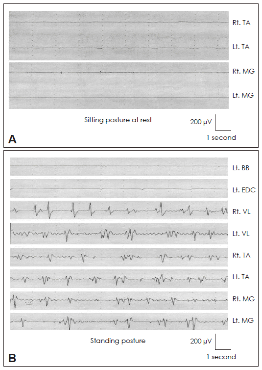
- None of any EMG bursts were observed on the both anterior tibialis (AT) and medial gastrocnemius (MG) as well as left biceps brachii and extensor digitorum communis muscles at sitting posture. While burst activities were recorded on the vastus lateralis, AT and MG muscle pairs of both legs for standing (Figure 1). The bursts length was 50 to 120 ms and frequencies variously ranged from 4 to 9 Hz with generally regular rhythms. The bursts were weakly presented on state, and more deteriorated on off state. Brain magnetic resonance imaging represented somewhat cerebral atrophy and old periventricular ischemia. Routine blood tests including a complete blood count, electrolytes, glucose level, renal function tests, liver function tests, and thyroid function tests showed no significant abnormal findings.
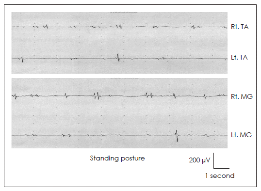
- When the levodopa was increased to 900 mg/day for 7 days by adding to existing treatment, standing movements in both legs were insidiously improved and jerky bursts of right leg on lying down were disappeared within 15 days. She could walk by herself for short distance (see Video, segment 2) and by cane for longer distance. She felt easier in standing still than before. Second followed EMG showed declined amplitude and frequencies of the burst movements (Figure 2).
Discussion
- Because of orthostatism and unsteadiness on standing posture, slow OT and OM is very difficult to be differentiated by visual analysis. Based on the surface electromyographic recordings in PD,6 slow-OT has lower frequency (4–9 Hz), more regular and longer bursts length (80–120 ms) than that of OM (9–15 Hz, irregular rhythm, 30–70 ms, respectively). EMG findings of our patient are characterized by lower frequencies (4–9 Hz), rhythm of moderate regularity, and burst synchrony or asynchrony with long length (50–120 ms). This is immediately distinguished from typical fast OT and seems similar to rest-tremor frequency range of patient. Rest tremor is frequently presented in slow-OT patients in the off state, whereas it rarely occur to the fast-OT or OM groups.6 Thus, orthostatism of our patient is near to slow OT, especially worsened on off state.
- The improvement of orthostatism after increasing levodopa also raises possibility of slow OT in this patient. This agreed with previous results of slow OT in PD with good response to levodopa.3,6 On the contrary, levodopa treatment might possibly be involved in the genesis of OM.6 Levodopa-induced myoclonus is usually associated with OM which is thought as other types of levodopa-induced dyskinesia.6 These considerations also suggest that orthostatism of our patient was not OM.
- OT plus with other neurological features is able to be related with abnormal dopaminergic pathway at the level of basal ganglia.2 And, this results in disinhibition of reticulospinal excitatory responses and causes an abnormal recruitment of spinal motor neurons.2 Therefore, we suggested that slow OT in PD may be at the least of partly responsible for the abnormal dopaminergic pathology.
- Legends to the Video
- Video Clip 1. The segment shows the involuntary jerks that predominantly involve the both legs. Myoclonus on both legs occurred with latency of 1 to 3 seconds after upright posture.
- Video Clip 2. After 1 week of treatment with increasing levodopa, the patient showed significant improvement of orthostatic myoclonus.
Acknowledgments
Acknowledgments
- This work was supported by Grant from Inje University, 2009.
Notes
-
The authors have no financial conflicts of interest.
Figure 1Surface electromyography (EMG) recordings of patient. First, there was no EMG bursts from both tibialis anterior (TA) and medial gastrocnemius (MG) muscles for sitting on a bed (A). Second, multichannel EMG recordings of a patient on standing showed brief irregular activities in the vastus lateralis, TA and MG muscles on both lower limbs in contrast to no discharge in the biceps brachii and extensor digitorum communis muscles on left upper limb (B).

Figure 2The electromyography recordings after increasing levodopa dosages were improved in both tibialis anterior and medial gastrocnemius muscles on standing.

REFERENCES
- 1. Deuschl G, Bain P, Brin M. Consensus statement of the Movement Disorder Society on Tremor. Ad Hoc Scientific Committee. Mov Disord 1998;13( Suppl 3):2–23.Article
- 2. Gerschlager W, Münchau A, Katzenschlager R, Brown P, Rothwell JC, Quinn N, et al. Natural history and syndromic associations of orthostatic tremor: a review of 41 patients. Mov Disord 2004;19:788–795.ArticlePubMed
- 3. Thomas A, Bonanni L, Antonini A, Barone P, Onofrj M. Dopa-responsive pseudo-orthostatic tremor in parkinsonism. Mov Disord 2007;22:1652–1656.ArticlePubMed
- 4. Apartis E, Tison F, Arné P, Jedynak CP, Vidailhet M. Fast orthostatic tremor in Parkinson’s disease mimicking primary orthostatic tremor. Mov Disord 2001;16:1133–1136.ArticlePubMed
- 5. Wills AJ, Brusa L, Wang HC, Brown P, Marsden CD. Levodopa may improve orthostatic tremor: case report and trial of treatment. J Neurol Neurosurg Psychiatry 1999;66:681–684.ArticlePubMedPMC
- 6. Leu-Semenescu S, Roze E, Vidailhet M, Legrand AP, Trocello JM, Cochen V, et al. Myoclonus or tremor in orthostatism: an under-recognized cause of unsteadiness in Parkinson’s disease. Mov Disord 2007;22:2063–2069.ArticlePubMed
- 7. Kim JS, Lee MC. Leg tremor mimicking orthostatic tremor as an initial manifestation of Parkinson’s disease. Mov Disord 1993;8:397–398.ArticlePubMed
- 8. Cristea RL, Goren H. Orthostatic tremor. Arch Neurol 1991;48:1119.ArticlePubMed
- 9. Glass GA, Ahlskog JE, Matsumoto JY. Orthostatic myoclonus: a contributor to gait decline in selected elderly. Neurology 2007;68:1826–1830.ArticlePubMed
Citations
Citations to this article as recorded by

- Orthostatic myoclonus – A retrospective study of Asian patients
Yi-Cheng Tai, Hung-Chang Kuo, Yihui Wu, Shih-Pin Hsu
Journal of the Formosan Medical Association.2022; 121(7): 1310. CrossRef - Therapeutic Devices for Motor Symptoms in Parkinson’s Disease: Current Progress and a Systematic Review of Recent Randomized Controlled Trials
Joji Fujikawa, Ryoma Morigaki, Nobuaki Yamamoto, Teruo Oda, Hiroshi Nakanishi, Yuishin Izumi, Yasushi Takagi
Frontiers in Aging Neuroscience.2022;[Epub] CrossRef - Unilateral Pseudo-Orthostatic Tremor Provoked by a Remote Limb Movement in Parkinson’s Disease
Sang-Won Yoo, Youngje Heo, Joong-Seok Kim, Kwang-Soo Lee
Journal of Movement Disorders.2020; 13(1): 69. CrossRef - Levodopa-Responsive Primary Slow Orthostatic Tremor: A Premotor Sign of Parkinson’s Disease?
Fumihito Yoshii, Wakoh Takahashi, Koji Aono
Case Reports in Neurology.2020; 12(1): 1. CrossRef


 KMDS
KMDS
 E-submission


 PubReader
 ePub Link
 Cite
Cite


