Articles
- Page Path
- HOME > J Mov Disord > Volume 3(1); 2010 > Article
-
Case Report
Restlessness with Manic Episodes due to Right Parietal Infarction - Suk Yun Kanga,b, Jong Won Paika, Young Ho Sohna
-
Journal of Movement Disorders 2010;3(1):22-24.
DOI: https://doi.org/10.14802/jmd.10007
Published online: April 30, 2010
aDepartment of Neurology and Brain Research Institute, Yonsei University College of Medicine, Seoul, Korea
bDepartment of Neurology, Kangnam Sacred Heart Hospital, Hallym University College of Medicine, Seoul, Korea
- Corresponding author Young Ho Sohn, MD, PhD, Department of Neurology and Brain Research Institute, Yonsei University College of Medicine, 250 Seongsan-ro, Seodaemun-gu, Seoul 120-752, Korea, Tel +82-2-2228-1601, Fax +82-2-393-0705, E-mail yhsohn62@yuhs.ac
• Received: October 16, 2009 • Accepted: April 21, 2010
Copyright © 2010 The Korean Movement Disorder Society
This is an Open Access article distributed under the terms of the Creative Commons Attribution Non-Commercial License (http://creativecommons.org/licenses/by-nc/3.0/) which permits unrestricted non-commercial use, distribution, and reproduction in any medium, provided the original work is properly cited.
- 14,991 Views
- 93 Download
- 4 Crossref
ABSTRACT
- Mood disorders following acute stroke are relatively common. However, restlessness with manic episodes has rarely been reported. Lesions responsible for post-stroke mania can be located in the thalamus, caudate nucleus, and temporal and frontal lobes. We present a patient who exhibited restlessness with manic episodes after an acute infarction in the right parietal lobe, and summarize the case reports involving post-stroke mania. The right parietal stroke causing mania in our case is a novel observation that may help us to understand the mechanisms underlying restlessness with mania following acute stroke.
- A 75-year-old right-handed woman was admitted because of the abrupt development of mental confusion, left hemiparesis, and dysarthria. She was previously healthy. One day before admission, she developed weakness in her left arm and disorientation, followed 20 hours later by inappropriate speech, agitation, and dysarthria. She had a history of hypertension and diabetes, but no history of psychiatric illness. She had no family history of affective illness.
- On admission, she showed partially impaired orientation to time, but had well-preserved orientation to place and person. The neurological examination revealed left homonymous hemianopsia, but no obvious motor or sensory deficits. She appeared agitated, and was talkative with an elated, euphoric mood. She also had a reduced need for sleep and pressured speech. She tended to touch the examiner while speaking. She showed no deficits in the line bisection and cancellation tasks, but was unable to copy an interlocked pentagon. She was also unable to draw a clock. Three days later, she developed motor weakness (grade 4) and hypesthesia in her left arm and face. Dysarthria developed along with persistence of her restlessness and manic features. The motor deficits and dysarthria were improved the next day, but the sensory deficit was unchanged.
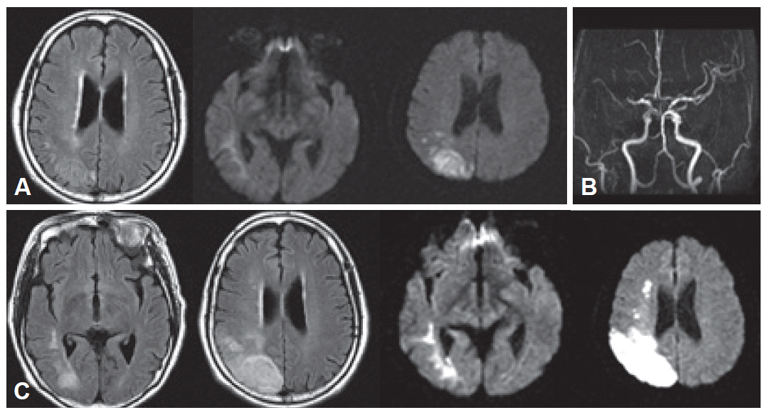
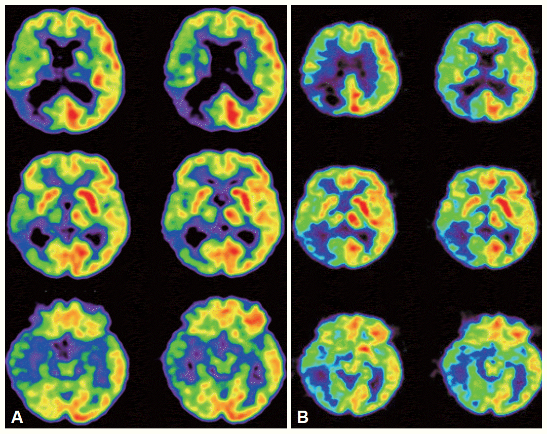
- Routine serologic testing and electrocardiography were all normal. Initial brain magnetic resonance imaging (MRI) with diffusion-weighted images (DWI) was performed on admission, which revealed an acute infarction in the right parietal lobe with subtle involvement of the posterior temporal area (Figure 1A). Magnetic resonance angiography (MRA) revealed an obstruction of portion M1 of the right middle cerebral artery (Figure 1B). Repeat MRI on the fourth hospital day after the motor deficits developed revealed that the infarction had increased in size (Figure 1C). Brain 2-deoxy-2-[18F]fluoro-D-glucose positron emission tomography (18F-FDG PET) performed 13 days after admission showed reduced FDG uptake in the right hemisphere, which was most severe in the right parietal lobe and left cerebellum (Figure 2A).
- When she was discharged 2 weeks after admission, her neurological deficits had improved. Her restlessness and manic symptoms had also partially alleviated without treatment. After 13 months, her neurological deficits and manic symptoms had resolved completely, except for the left-sided visual field defect. Repeated brain 18F-FDG PET showed that the previous reduction in FDG uptake in the right hemisphere had improved (Figure 2B).
Case
- This patient had sudden-onset restlessness with manic symptoms, including agitated mood, irritability, euphoria, talkativeness, pressured speech, and decreased need for sleep. The close temporal relationship between the onset of her symptoms and ischemic stroke, along with the absence of a history of affective disorders, supports the hypothesis that her restlessness with mania was caused by acute infarction of the right parietal lobe.
- An extensive literature search revealed 17 reports that described 29 patients who had post-stroke manic episodes. Of these, eleven patients developed manic symptoms concomitantly with their strokes. In the other cases, the temporal relationship between the stroke and mania was obscure, either with a fairly long latency or due to insufficient information. The lesions were located mostly in the right hemisphere. Only five patients had left-sided lesions. The reported anatomical locations of the strokes were the temporal and frontal cortex (13 patients), 1,2,5,6,15–17 thalamus (9 patients),3,5,8,9,11,13 caudate nucleus (2 patients),6 and ventral pontine region (2 patients).7 Some patients had infarctions in widespread regions involving the frontal, temporal, and parietal lobes. Our patient had an infarction that was mainly confined to the right parietal region on the initial brain MRI, although there was subtle involvement in the posterior temporal area. Post-stroke mania due to a localized infarction in the right parietal lobe has not previously been reported. One report described a case affecting the right parieto-occipital area involving the geniculocalcarine tract with agitation, hallucinations, and delusion, but the computed tomography result was not reported.18 Widespread reduction in right hemispheric activity was seen on the brain FDG PET. Although there was no structural damage (i.e., infarction), this may have been responsible for the restlessness and manic symptoms in this patient. However, the persistence of reduced activity in the right hemisphere, as shown on the repeated brain PET, along with a complete resolution of the manic symptoms, supports the possible relationship between the manic symptoms and the right parietal infarction.
- Disinhibition syndrome, including restlessness and manic features, is usually caused by lesions in the orbitofrontal and basotemporal cortices of the right hemisphere.5,19 These areas are thought to inhibit the motor, instinctive, affective, and intellectual behaviors elaborated in the dorsal cortex selectively.19 Lesions in these areas or in their connections to the dorsal cortex could produce disinhibition syndrome. This may range from mildly inappropriate social behavior to full-blown mania.19 Caplan et al.20 observed that about half of the patients with infarcts of the inferior division of the right middle cerebral artery were in an agitated, confused state, showing hyperactivity, restlessness, and easy distractibility. Their motor and sensory deficits were usually mild and transient. These features are consistent with those of our patient. As Caplan et al.20 suggested, the inferior parietal lesions in our patient may interrupt the connections between the medial limbic cortex and superior parietal and frontal lobes, altering the emotional tone and affective behavior.
Discussion
Figure 1A: Brain MRI on admission shows an acute right parietal lobe infarction on FLAIR (left) and diffusion-weighted images (middle, right). B: Brain MRA shows obstruction of portion M1 of the right middle cerebral artery. C: Three days after admission, new neurological deficits developed. Follow-up brain MRI shows the infarct is larger.


Figure 2A: Brain 18F-FDG PET 13 days after admission shows diffusely decreased FDG uptake in the right hemisphere, most severely affecting the right parietal area. The FDG uptake in the right basal ganglia and thalamus is also decreased. B: At 13 months, repeat 18F-FDG PET shows improved FDG uptake in the right hemisphere, compared to the initial PET. 18F-FDG PET: [18F] fluoro-D-glucose prositron emission tomography.


- 1. Cohen MR, Niska RW. Localized right cerebral hemisphere dysfunction and recurrent mania. Am J Psychiatry 1980;137:847–848.ArticlePubMed
- 2. Jampala VC, Abrams R. Mania secondary to left and right hemisphere damage. Am J Psychiatry 1983;140:1197–1199.ArticlePubMed
- 3. Cummings JL, Mendez MF. Secondary mania with focal cerebrovascular lesions. Am J Psychiatry 1984;141:1084–1087.ArticlePubMed
- 4. Bogousslavsky J, Ferrazzini M, Regli F, Assal G, Tanabe H, Delaloye-Bischof A. Manic delirium and frontal-like syndrome with paramedian infarction of the right thalamus. J Neurol Neurosurg Psychiatry 1988;51:116–119.ArticlePubMedPMC
- 5. Starkstein SE, Boston JD, Robinson RG. Mechanisms of mania after brain injury. 12 case reports and review of the literature. J Nerv Ment Dis 1988;176:87–100.ArticlePubMed
- 6. Starkstein SE, Mayberg HS, Berthier ML, Fedoroff P, Price TR, Dannals RF, et al. Mania after brain injury: neuroradiological and metabolic findings. Ann Neurol 1990;27:652–659.ArticlePubMed
- 7. Drake ME Jr, Pakalnis A, Phillips B. Secondary mania after ventral pontine infarction. J Neuropsychiatry Clini Neurosci 1990;2:322–325.Article
- 8. Kulisevsky J, Berthier ML, Pujol J. Hemiballismus and secondary mania following a right thalamic infarction. Neurology 1993;43:1422–1424.ArticlePubMed
- 9. Vuilleummier P, Ghika-Schmid F, Bogousslavsky J, Assal G, Regli F. Persistent recurrence of hypomania and prosopoaffective agnosia in a patient with right thalamic infarct. Neuropsychiatry Neuropsychol Behav Neurol 1998;11:40–44.PubMed
- 10. Fenn D, George K. Post-stroke mania late in life involving the left hemisphere. Aust N Z J Psychiatry 1999;33:598–600.ArticlePubMed
- 11. Leibson E. Anosognosia and mania associated with right thalamic haemorrhage. J Neurol Neurosurgery Psychiatry 2000;68:107–108.ArticlePubMedPMC
- 12. Benke T, Kurzthaler I, Schmidauer Ch, Moncayo R, Donnemiller E. Mania caused by a diencephalic lesion. Neuropsychologia 2002;40:245–252.ArticlePubMed
- 13. Huffman J, Stern TA. Acute psychiatric manifestations of stroke: a clinical case conference. Psychosomatics 2003;44:65–75.ArticlePubMed
- 14. Celik Y, Erdogan E, Tuglu C, Utku U. Post-stroke mania in late life due to right temporoparietal infarction. Psychiatry Clin Neurosci 2004;58:446–447.ArticlePubMed
- 15. Mimura M, Nakagome K, Hirashima N, Ishiwata H, Kamijima K, Shinozuka A, et al. Left frontotemporal hyperperfusion in a patient with post-stroke mania. Psychiatry Res 2005;139:263–267.ArticlePubMed
- 16. Goyal R, Sameer M, Chandrasekaran R. Mania secondary to right-sided stroke--responsive to olanzapine. Gen Hosp Psychiatry 2006;28:262–263.ArticlePubMed
- 17. Nagaratnam N, Wong KK, Patel I. Secondary mania of vascular origin in elderly patients: a report of two clinical cases. Arch Gerontol Geriatr 2006;43:223–232.ArticlePubMed
- 18. Pakalnis A, Drake ME Jr, Kellum JB. Right parieto-occipital lacunar infarction with agitation, hallucinations, and delusions. Psychosomatics 1987;28:95–96.ArticlePubMed
- 19. Starkstein SE, Robinson RG. Mechanism of disinhibition after brain lesions. J Nerv Ment Dis 1997;185:108–114.ArticlePubMed
- 20. Caplan LR, Kelly M, Kase CS, Hier DB, White JL, Tatemichi T, et al. Infarcts of the inferior division of the right middle cerebral artery: mirror image of Wernicke’s aphasia. Neurology 1986;36:1015–1020.ArticlePubMed
REFERENCES
Figure & Data
References
Citations
Citations to this article as recorded by 

- Restlessness with manic episodes induced by right-sided multiple strokes after COVID-19 infection: A case report
Takahiko Nagamine
Brain Circulation.2023; 9(2): 112. CrossRef - Poststroke Mania During the COVID-19 Pandemic
Takahiko Nagamine
Journal of Nervous & Mental Disease.2023; 211(12): 979. CrossRef - Management of psychiatric disorders in patients with stroke and traumatic brain injury
Gautam Saha, Kaustav Chakraborty, Amrit Pattojoshi
Indian Journal of Psychiatry.2022; 64(8): 344. CrossRef - Post stroke delirium
M. A. Savina
Zhurnal nevrologii i psikhiatrii im. S.S. Korsakova.2014; 114(12. Vyp. 2): 19. CrossRef
Comments on this article
 KMDS
KMDS
 E-submission
 PubReader
 ePub Link
 Cite
Cite


